The knee joint consists of a network of soft tissues, including cartilage, tendon, and ligaments, that provide flexibility, stability, and strength to the joint.
In This Article:
- Guide to Knee Joint Anatomy
- Soft Tissue of the Knee Joint
- How Knee Joint Problems Cause Pain
Cartilage of the Knee Joint
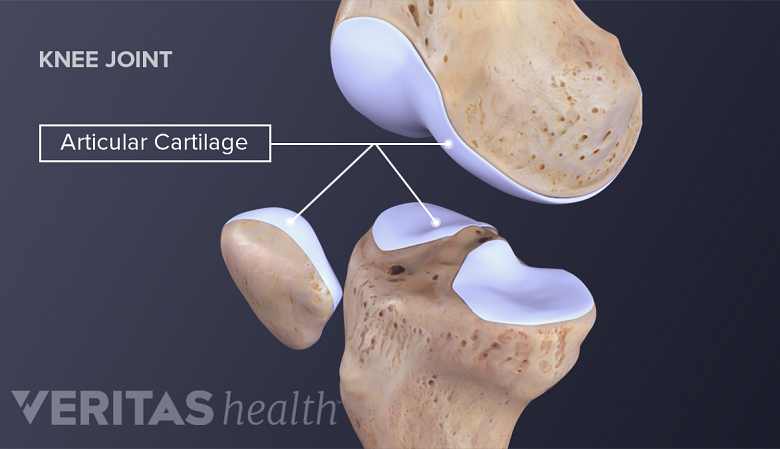
Articular cartilage allows knee bones to move over one another with minimal friction.
Slippery and flexible, hyaline (articular) cartilage within the knee joint allows, has less friction than two pieces of glass placed together. This allows the joint to move with minimal friction in a healthy knee. There are two primary types of cartilage in the knee:
- Articular (Hyaline) cartilage. This cartilage covers the bones where they meet at the knee joint. The end of the femur (condyles) and the back of the patella (knee cap). It is simultaneously smooth and strong, allowing bones to move over one another with minimal friction.
- Meniscus (Fibrocartilage). The menisci are two C-shaped cartilaginous structures within the knee. The medial (inside) and lateral (outer) menisci act as cushions for weight bearing activities, decreasing the effect of impact between the femur and tibia.
The articular cartilage and menisci are integral to the proper function of the knee joint. They can be injured in an acutely or gradually through repetitive stresses. The loss of cartilage in the knee is termed arthritis. This can be both inflammatory, as in rheumatoid arthritis, or primary, as in the case of osteoarthritis.
Tendons, Ligaments, and Other Soft Tissues of the Knee Joint
The knee joint relies on a variety of ligaments, tendons, and soft tissue structures to maintain flexibility, stability, and strength.
Ligaments are ropy, fibrous bands of tissue that connect bones to other bones.
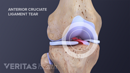
- The Anterior Cruciate Ligament (ACL). The ACL connects the tibia to the femur and functions to prevent the tibia from sliding forward on the femur. The ACL is commonly injured in sporting activities and rarely injured in isolation.
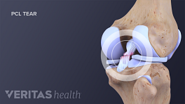
- The Posterior Cruciate Ligament (PCL). The PCL also connects the tibia to the femur. It functions to prevent the tibia from sliding backward on the femur. The PCL works with the ACL for stabilization of the knee. It is commonly injured in hyperextension type knee moments.
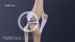
- The Lateral Collateral Ligament (LCL). The LCL, which is also known as the fibular collateral ligament, is located on the outside (lateral side) of the knee. It connects the outside, bottom edge of the femur to the outside, top edge of the fibula. The LCL helps stabilize the knee joint by limiting outward (varus) force across the knee.
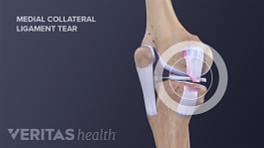
- The Medial Collateral Ligament (MCL). The MCL is located on the inside (medial side) of the knee, connecting the inside, bottom edge of the femur with the inside, top edge of the tibia. The MCL helps to stabilize the knee by limiting inward (valgus) force across the knee. The MCL works with the LCL to prevent unwanted side-to-side motion. The MCL is the most commonly injured knee ligament.
Tendons are flexible tissues that attach muscle to bone.
- The hamstring tendons. There are three hamstring tendons that cross the knee joint on the back of the knee. Two are on the inside (medial) part of the knee attaching to the shin bone (Semimembranosus and Semitendinosus) and one is on the outside (lateral) part of the knee, attaching to the fibula (Biceps femoris).
See Symptoms of Chronic High (Proximal) Hamstring Tendinopathy
- The quadriceps tendon. This tendon is composed of contributions from the four quadriceps muscles (the vastus lateralis, vastus intermedius, vastus medialis, and rectus femoris). It attaches the powerful quadriceps muscles to the top part of the patella.
- The patellar tendon. This tendon (also called patellar ligament) attaches the bottom part of the patella to the top part of the tibia.
- A synovial membrane. All of the joints in the body are surrounded by a balloon that holds the joint fluid in. These balloons hold the joint fluid (synovial fluid) in the joint. This fluid is integral to the health of the joint, providing lubrication and delivering nutrients.