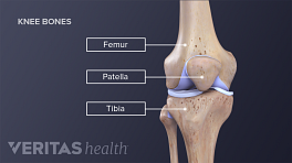
Before diagnosing a medial collateral ligament (MCL) injury, a health care professional must rule out the possibility of other knee injuries or conditions, such as a meniscus tear. Getting an accurate diagnosis will begin with an in-office doctor evaluation.
In This Article:
Doctor Evaluation
Generally, a health care professional can make a diagnosis of an MCL tear through a detailed patient interview and careful physical examination.
Patient interview
A person should be prepared to answer questions, such as:
- How did the injury occur?
- When did the injury occur?
- Where is the location of the pain?
- If swelling is present, when did it start?
- Was the trauma accompanied by a popping or tearing sensation?
- Has the knee been injured before?
Physical examination
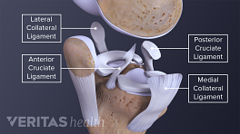
During a physical examination a doctor will likely check for pain and tenderness in the affected knee. Additionally, the patient may go through a series of range-of-motion tests to determine if the MCL is injured and whether or not other injuries are present, such as an ACL or meniscal tear.
The most common exam is the valgus stress test. The doctor will apply outside force to the injured knee at certain angles. If there is pain, then the MCL may be injured.
Diagnostic Imaging
A doctor may order one or more medical imaging tests to confirm the presence and determine the severity of an MCL injury.
- X-rays use low levels of radiation and give doctors a view of a person’s bones. Although MCL injuries do not show up on standard X-ray exams, they are a relatively inexpensive, fast way to rule out other possible injuries that might be causing the symptoms. Additionally, a stress X-ray—where a physician applies a valgus force to the knee during the exam—can help to determine the degree of ligamentous injury.
- Magnetic resonance imaging (MRI) shows a detailed view of the soft tissue surrounding the knee joint. An MRI can also help a doctor determine the location and grade of an MCL tear.
- Ultrasound imaging uses high-frequency sound waves to build a picture of the knee’s tissues. Ultrasound can be utilized in situations when an MRI is not recommended. Ultrasound may also be used in an urgent care setting to make an immediate assessment, allowing the injury to be treated more quickly. 1 Ghosh N, Kruse D, Subeh M, Lahham S, Fox JC. Comparing Point-of-care-ultrasound (POCUS) to MRI for the Diagnosis of Medial Compartment Knee Injuries. J Med Ultrasound. 2017;25(3):167–172. doi:10.1016/j.jmu.2017.06.004
Diagnostic imaging is not always required to diagnose an MCL injury.
Once a person is diagnosed, treatment options can be recommended.
- 1 Ghosh N, Kruse D, Subeh M, Lahham S, Fox JC. Comparing Point-of-care-ultrasound (POCUS) to MRI for the Diagnosis of Medial Compartment Knee Injuries. J Med Ultrasound. 2017;25(3):167–172. doi:10.1016/j.jmu.2017.06.004