
Subscribe to our newsletter and get your FREE guide,
Natural Back Pain Relief: 16 Choices for Lasting Comfort.
Stress fractures, sometimes called hairline fractures, often occur in the feet, because of the weight-bearing responsibility of these bones. Stress fractures are small, microscopic cracks in bone that occur when the bone is unable to handle the load, or weight, placed on it.
Foot stress fractures are often caused by overuse or repetitive activities. Because of this, stress fractures are commonly seen in athletes, such as runners, soccer players, or dancers, but they can also be seen in normal people who have changed their daily activities.
See All About Stress Fractures
Foot stress fractures also occur more frequently in people with health problems that affect bones such as osteoporosis, or people with abnormal gait or other problems, such as bunions or tendonitis.
Overuse or repetitive injury is a common cause of stress fractures in the foot.
In This Article:
Mechanism of Injury: Foot Stress Fractures
Bones are living tissues that are constantly adapting to loads by rebuilding and repairing. This process is called remodeling and is the reason that bones can heal after injury. This process also helps bones become stronger when loaded appropriately, and the reason bones become weaker if they are not exposed to sufficient loads.1Mandell JC, Khurana B, Smith SE. Stress fractures of the foot and ankle, part 1: biomechanics of bone and principles of imaging and treatment. Skeletal Radiol. 2017 Aug;46(8):1021-1029. doi: 10.1007/s00256-017-2640-7. Epub 2017 Apr 4.
advertisement
Stress fractures occur when the loading on the bone is greater than the bone’s ability to adapt to stress. Before the bone fractures, there can be pain in the area due to swelling and abnormal remodeling. This is termed a stress reaction, and can be seen on specialized imaging.
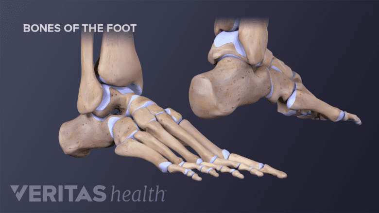
Foot Anatomy: Where Stress Fractures Occur

Stress fractures can occur in any bone of the foot, however, certain bones are affected more often than others.
- The metatarsal bones, which connect the toes to the midfoot. The second and third metatarsal, which connect the second and third toe, are most commonly injured, and represent 17-35% of all stress fractures of the foot and ankle.2Mandell JC, Khurana B, Smith SE. Stress fractures of the foot and ankle, part 2: site-specific etiology, imaging, and treatment, and differential diagnosis. Skeletal Radiol. 2017 Sep;46(9):1165-1186. doi: 10.1007/s00256-017-2632-7. Epub 2017 Mar 25. Metatarsal fractures are common in dancers and runners.3Mayer SW, Joyner PW, Almekinders LC, Parekh SG. Stress fractures of the foot and ankle in athletes [published correction appears in Sports Health. 2015 Nov;7(6):557]. Sports Health. 2014;6(6):481–491. doi:10.1177/1941738113486588
- The calcaneus bone, also called the heel bone, forms the foundation of the back of the foot. The calcaneus bone represents approximately 21-28% of stress fractures of the foot and ankle.1Mandell JC, Khurana B, Smith SE. Stress fractures of the foot and ankle, part 1: biomechanics of bone and principles of imaging and treatment. Skeletal Radiol. 2017 Aug;46(8):1021-1029. doi: 10.1007/s00256-017-2640-7. Epub 2017 Apr 4.,2Mandell JC, Khurana B, Smith SE. Stress fractures of the foot and ankle, part 2: site-specific etiology, imaging, and treatment, and differential diagnosis. Skeletal Radiol. 2017 Sep;46(9):1165-1186. doi: 10.1007/s00256-017-2632-7. Epub 2017 Mar 25.
- The navicular bone, which sits near the top of the foot.
advertisement
In children, stress fractures in the foot can occur in areas of bone growth. Common locations for foot stress fractures in children include the calcaneus, cuboid, talus, and navicular bone.4Oestreich AE, Bhojwani N. Stress fractures of ankle and wrist in childhood: nature and frequency. Pediatr Radiol. 2010 Aug;40(8):1387-9. doi: 10.1007/s00247-010-1577-y. Epub 2010 Feb 24.
The anatomical location of the stress fracture determines how it heals. Stress fractures in areas of tensile forces or areas with poor blood flow do not heal as well as stress fracture in areas with compressive forces and good blood flow.1Mandell JC, Khurana B, Smith SE. Stress fractures of the foot and ankle, part 1: biomechanics of bone and principles of imaging and treatment. Skeletal Radiol. 2017 Aug;46(8):1021-1029. doi: 10.1007/s00256-017-2640-7. Epub 2017 Apr 4.
- 1 Mandell JC, Khurana B, Smith SE. Stress fractures of the foot and ankle, part 1: biomechanics of bone and principles of imaging and treatment. Skeletal Radiol. 2017 Aug;46(8):1021-1029. doi: 10.1007/s00256-017-2640-7. Epub 2017 Apr 4.
- 2 Mandell JC, Khurana B, Smith SE. Stress fractures of the foot and ankle, part 2: site-specific etiology, imaging, and treatment, and differential diagnosis. Skeletal Radiol. 2017 Sep;46(9):1165-1186. doi: 10.1007/s00256-017-2632-7. Epub 2017 Mar 25.
- 3 Mayer SW, Joyner PW, Almekinders LC, Parekh SG. Stress fractures of the foot and ankle in athletes [published correction appears in Sports Health. 2015 Nov;7(6):557]. Sports Health. 2014;6(6):481–491. doi:10.1177/1941738113486588
- 4 Oestreich AE, Bhojwani N. Stress fractures of ankle and wrist in childhood: nature and frequency. Pediatr Radiol. 2010 Aug;40(8):1387-9. doi: 10.1007/s00247-010-1577-y. Epub 2010 Feb 24.

advertisement