
Subscribe to our newsletter and get your FREE guide,
Natural Back Pain Relief: 16 Choices for Lasting Comfort.
There are many different muscles and ligaments in the ankle, which gives the ankle its strength, flexibility, and range of motion.
In This Article:
Major ligaments of the ankle

Important ligaments located in the front of the ankle.
Ligaments are a type of soft tissue that is made up mostly of collagen. Ligaments have low vascularity, which means they do not receive much blood flow. This lack of blood flow makes ligaments slower to heal than other types of soft tissue.
Unlike tendons, which connect muscle to bone, ligaments connect bones to other bones.
advertisement
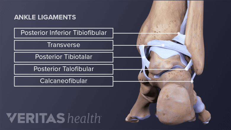
Important ligaments located at the back of the ankle.
There are several major ligaments in the ankle:
- Three ligaments on the outside of the ankle that make up the lateral ligament complex, as follows:
- The anterior talofibular ligament (ATFL), which connects the front of the talus bone to a long bone in the lower leg called the fibula
- The calcaneofibular ligament (CFL), which connects the calcaneus, or heel bone, to the fibula
- The posterior talofibular ligament (PTFL), which connects the rear of the talus bone to the fibula
- The deltoid ligament, a thick ligament that supports the entire medial, or inner, side of the ankle and is made up of parts:
- Anterior tibiotalar ligament (ATTL)
- Posterior tibiotalar ligament (PTTL)
- Tibiocalcaneal ligament (TCL)
- Tibionavicular ligament (TNL)
- The anterior inferior tibiofibular ligament (AITFL), which connects the tibia to the fibula
- Two posterior fibular ligaments, which crisscross the back of the tibia and fibula:
- The posterior inferior tibiofibular ligament (PITFL)
- The transverse ligament
- The interosseous ligament, which rests between the tibia and fibula and runs the entire length of the tibia and fibula, from the ankle to the knee
The various ligaments that surround the ankle together help form part of the joint capsule, a fluid-filled sac that surrounds and lubricates articulating joints.
advertisement
Major muscles of the ankle
There are also multiple muscles in the ankle that can be strained, as follows:
- The peroneal muscles (peroneus longus and peroneus brevis), on the outside edge of the ankle and foot. These muscles allow the ankle to bend downward and outward.
- The calf muscles (gastrocnemius and soleus), which are connected to the calcaneus via the Achilles tendon. The tightening and relaxing of the calf muscles enables the ankle to bend downward and upward.
- The posterior tibialis muscle, which supports the arch of the foot and enables the foot to turn inward.
- The anterior tibialis muscle, which enables the ankle and foot to turn upward.1Sechrest, R. Ankle anatomy: a patient's guide. eOrthopod.com: http://www.eorthopod.com/ankle-anatomy/topic/159. Accessed November 17, 2014.
The complexity of the ankle's muscular and ligament structure creates many possible opportunities for injuries when the ankle is pushed beyond its normal range of motion.
- 1 Sechrest, R. Ankle anatomy: a patient's guide. eOrthopod.com: http://www.eorthopod.com/ankle-anatomy/topic/159. Accessed November 17, 2014.

advertisement