
Subscribe to our newsletter and get your FREE guide,
Natural Back Pain Relief: 16 Choices for Lasting Comfort.
A proximal humeral fracture is diagnosed if there is a bone break in the area surrounding the humeral head, commonly known as the ball of the shoulder’s ball-and-socket joint. The humeral head is located at the top of the humerus (upper arm bone).

Proximal humerus fractures occur at or immediately below the “ball” of the shoulder joint.
In This Article:
- Proximal Humerus Fractures of the Shoulder
- Treating a Proximal Humerus Fracture
The Essential Things to Know About a Proximal Humerus Fracture
The proximal humerus is the part of the upper arm bone that forms a major part of the shoulder joint.
- Proximal humerus fractures are also called fragility fractures and are common in people over 65 years of age who have weakened bones due to osteoporosis.1Pencle F, Varacallo M. Proximal Humerus Fracture. [Updated 2023 Aug 4]. In: StatPearls [Internet]. Treasure Island (FL): StatPearls Publishing; 2024 Jan-. Available from: https://www.ncbi.nlm.nih.gov/books/NBK470346/
- The fracture can involve the “ball” of the upper arm bone and/or the part of bone below the spherical portion.
- A proximal humerus fracture generally causes severe pain and greatly affects the shoulder joint and upper arm movement. See Bone Break vs. Fracture
- Most people recover well with medical treatment and regain normal function, but some individuals experience ongoing limitations.
Proximal humerus fractures are among the 3 common types of bone fracture in people with osteoporosis along with vertebral fractures and distal radius (wrist) fractures.1Pencle F, Varacallo M. Proximal Humerus Fracture. [Updated 2023 Aug 4]. In: StatPearls [Internet]. Treasure Island (FL): StatPearls Publishing; 2024 Jan-. Available from: https://www.ncbi.nlm.nih.gov/books/NBK470346/
advertisement
Types
Doctors use one of three classification systems when evaluating a proximal humerus fracture.
Neer classification
In this system, the proximal humerus is divided into four parts, and then, the fracture is classified based on the number and position of the broken bone pieces:
One-part fracture
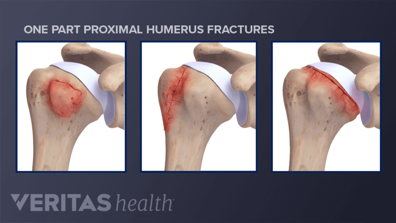
In a one-part fracture, the bone is broken in one to four places, and all the fragments remain aligned in their original position.
About ¾ of all proximal humerus fractures fall into this classification and most can be treated without surgery.
Two-part fracture

In this fracture type, the bone is broken in two to four places, and one fragment is displaced.
This fracture type typically accounts for most of the remaining ¼ of proximal humerus fractures.
Three-part fracture

In a three-part fracture, the bone is broken in three to four places, with two fragments displaced.
Four-part fracture
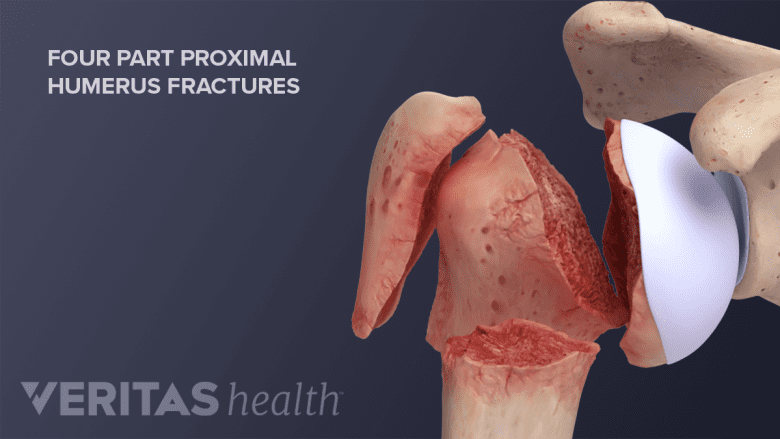
Four-part fracture comprises bone broken in more than four places, with three fragments displaced.
A fragment is considered displaced when it has moved more than 1 cm or more than 45 degrees from its original position.1
As discussed above, the majority of proximal humeral fractures are non-displaced, meaning the broken bones remain in their correct anatomical position.
AO classification
This classification system is based on the complexity of the fracture. It provides a detailed framework for understanding the fracture pattern and guiding treatment decisions.
The AO classification divides proximal humerus fractures into three main types:
- Type A: extra-articular, unifocal, or two-part fractures
- Involves a single fracture line without affecting the joint surface.
- Type B: extra-articular, bifocal, or three-part fractures
- Involves two fracture lines without affecting the joint surface.
- Type C: multifocal, four-part, or articular fractures
- Involves multiple fracture lines and often affects the joint surface.
Each type is further classified based on specific fracture patterns and characteristics, allowing for more precise classification and treatment planning.
Fracture type based on the anatomical location
Proximal humerus fractures are also classified based on the part of the bone that is affected:
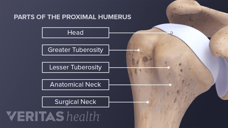
- Greater tuberosity fracture: a break in the bony prominence at the top, outer portion of the humerus.
- Lesser tuberosity fracture: a break in the smaller bony prominence at the top, inner portion of the humerus.
- Surgical neck fracture: a break in the narrow part of the humerus just below the “ball.”
- Anatomical neck fracture: a break in the “ball” or joint surface of the proximal humerus.
These are general categories, and there can be variations and combinations of these fracture types.
Symptoms and Signs

It is common to experience shoulder pain, swelling, bruising, and weakness after a proximal humerus fracture.
The fracture typically causes some combination of these symptoms:
- Sharp, intense pain in the shoulder area, which may travel down the arm.
- Inflammation and swelling around the shoulder joint.
- Bruising of the skin, often extending from the shoulder down to the elbow, due to bleeding beneath the surface.
- A noticeable lump or deformity in the shoulder area.
- Shoulder pain when moving the arm, especially while lifting, lowering, or rotating the arm.
- Grinding or crunching sensation when moving the shoulder joint, caused by the fractured bone ends rubbing together.
- Numbness or tingling in the affected arm or hand, caused by nerve damage.
- Weakness and loss of strength in the affected arm.
Shoulder pain and discomfort often make it difficult to find a comfortable sleeping position.
advertisement
Causes
Proximal humerus fractures commonly occur due to a fall on the shoulder or falling on an outstretched hand.
- In older individuals, the fracture occurs due to low-energy falls, such as losing balance while walking, running, or climbing stairs.
- In younger individuals, the fracture occurs due to high-intensity mechanisms, such as:
- Falling from a two-wheeled vehicle
- Falls during athletic events, such as biking, horseback riding, and skateboarding
- Contact sports, such as football, rugby, and hockey
A less common cause of proximal humerus fractures and dislocations is intense muscle contractions due to seizure or electric shock, which occurs more commonly in the younger population.
Diagnosis
A proximal humerus fracture is typically diagnosed through a combination of:
- A medical history review, which is crucial in identifying potential causes such as falls
- A physical examination
- An x-ray, which usually confirms the diagnosis
Physical exam
A thorough physical examination is conducted where the doctor examines the shoulder for:
- Swelling
- Tenderness
- Limitations in movement
- Neurological symptoms, such as numbness or tingling
The doctor also palpates (gently feels) the shoulder blade and ribs to determine if there is an accompanying injury.
A neurological examination is performed to evaluate nerve functions in the neck, arm, and hand.
X-ray
The primary imaging test for proximal humerus fractures is an x-ray. Multiple views of the shoulder are taken to assess the severity and location of the break.
X-rays are also used to track bone healing progress during follow-up appointments.
For severe fractures or if other injuries are suspected, CT or MRI scans are used.
Computed tomography (CT)
A CT scan provides detailed images of the bone and surrounding soft tissues. It is often used to assess the complexity of the fracture and plan surgical treatment.
Magnetic resonance imaging (MRI)
An MRI provides detailed information about soft tissue damage, including cartilage, tendons, and ligaments. It is used when there is a suspicion of nerve or blood vessel injury.
advertisement
How Long Does a Fractured Proximal Humerus Take to Heal?
A simple, uncomplicated fracture typically heals in about six weeks.
Healing times are extended in cases such as complex fractures, advanced age, smoking/nicotine use, and/or the presence of underlying conditions such as osteoporosis or diabetes. In these cases, proximal humerus fractures can take several months to fully heal and regain strength in the arm.
Once the initial inflammation has reduced, guided physical therapy is recommended to strengthen the shoulder muscles and aid in overall recovery.
Accelerating the healing process of a fracture involves a combination of medical care and self-care. Several factors contribute to optimal bone repair and significantly impact recovery time, including immobilizing the broken bone, getting sufficient rest, engaging in prescribed exercises, and consuming a balanced diet rich in bone-building nutrients.
When to See a Doctor
Prompt evaluation and treatment of a proximal humerus fracture ensures adequate bone healing and prevents complications such as nonunion of the fractured ends.
A fracture following a traumatic event or a suspected fracture with the following symptoms and signs warrants immediate medical attention:
- Break in the skin or tenting (cone-shaped bulge) of the skin with visible bone protrusion
- Neurologic deficits such as numbness or weakness in the arm or hand on the affected side
- Drooping or sagging shoulder
- Severely swollen shoulder
- Severe bleeding
- Difficulty breathing
- Chest pain
While waiting to see a doctor, patients should immobilize the injured arm and shoulder by holding it close to their body with their other arm.
Proximal humerus fractures are diagnosed and treated by orthopedic surgeons and sports medicine specialists. These physicians often coordinate care with physical therapists and primary care doctors.
- 1 Pencle F, Varacallo M. Proximal Humerus Fracture. [Updated 2023 Aug 4]. In: StatPearls [Internet]. Treasure Island (FL): StatPearls Publishing; 2024 Jan-. Available from: https://www.ncbi.nlm.nih.gov/books/NBK470346/

advertisement