
Subscribe to our newsletter and get your FREE guide,
Natural Back Pain Relief: 16 Choices for Lasting Comfort.
The soft tissues surrounding the shoulder joints provide strength, stability, and resilience to the joint in the resting position and while performing different movements.
In This Article:
- Guide to Shoulder Anatomy
- Soft Tissues of the Shoulder
Soft Tissues Involved in Joint Stability: Capsule, Bursae, Labrum, and Ligaments
The soft tissues that help to maintain the stability of the shoulder joints at rest include:
Joint capsule

The joint capsule provides stability and cushioning to the joint.
The joint capsule envelopes the glenohumeral joint – a ball and socket joint that connects the round head of the upper arm (humerus) bone to a shallow cavity in the shoulder blade (glenoid cavity).1Chang LR, Anand P, Varacallo M. Anatomy, Shoulder and Upper Limb, Glenohumeral Joint. In: StatPearls. StatPearls Publishing; 2023. Accessed July 27, 2023. http://www.ncbi.nlm.nih.gov/books/NBK537018/ The joint capsule is composed of dense fibrous connective tissue and provides stability to the glenohumeral joint at rest and during movements.
A synovial membrane (specialized connective tissue) lines the inner surface of the joint capsule and secretes synovial fluid. The synovial fluid lubricates the head of the upper arm bone and the surface of the glenoid cavity.
A similar type of synovial fluid-filled joint capsule also provides stability and cushioning to the acromioclavicular joint (joint between the shoulder blade and collar bone).2Wong M, Kiel J. Anatomy, Shoulder and Upper Limb, Acromioclavicular Joint. In: StatPearls. StatPearls Publishing; 2023. Accessed July 28, 2023. http://www.ncbi.nlm.nih.gov/books/NBK499858/
Bursae
Bursae provide cushioning to the bones.
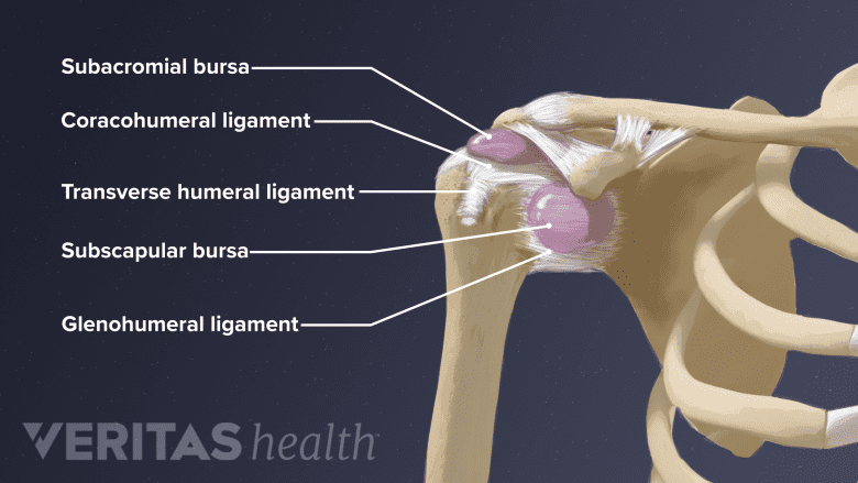
In addition to the joint capsule, the glenohumeral joint is also surrounded by synovial fluid-filled sacs called the bursae. The bursae help cushion the joint and reduce friction between the bones, muscles, tendons, and ligaments that meet at the glenohumeral joint. The bursae surrounding the glenohumeral joint include3Mostafa E, Imonugo O, Varacallo M. Anatomy, Shoulder and Upper Limb, Humerus. In: StatPearls. StatPearls Publishing; 2023. Accessed July 28, 2023. http://www.ncbi.nlm.nih.gov/books/NBK534821/:
- The subacromial bursa
- The subdeltoid bursa
- The subscapular bursa and
- The subcoracoid bursa
Overuse of the shoulder or a tear in the surrounding tendons can cause inflammation of the bursa in the shoulder. An inflammation of the bursa is known as bursitis.
Read more about Shoulder Bursitis on Arthritis-health.com.
For instance, inflammation of the subacromial bursa (subacromial bursitis) is a common cause of shoulder pain. Subacromial bursitis is typically observed in individuals who frequently engage in overhead activities,4Faruqi T, Rizvi TJ. Subacromial Bursitis. In: StatPearls [Internet]. Treasure Island (FL): StatPearls Publishing; 2023 Jan. Accessed August 28, 2023. https://www.ncbi.nlm.nih.gov/books/NBK541096/ such as:
- Sports like tennis, volleyball, swimming, and baseball
- Physically demanding jobs like wall painting, hairdressing, and carpentry
Another common cause of pain in the shoulder region is the inflammation of the bursae in the shoulder blades, known as the scapulothoracic bursae. The scapulothoracic bursae include the infraserratus, subserratus, and trapezoid bursae, which cushion the movement between the shoulder blade and the rib cage wall. The inflammation of any of these scapulothoracic bursae results in a condition called snapping scapula syndrome.5Conduah AH, Baker CL, Baker CL. Clinical Management of Scapulothoracic Bursitis and the Snapping Scapula. Sports Health. 2010;2(2):147-155. doi:10.1177/1941738109338359
Labrum
The shoulder’s labrum is a fibrous and rigid cartilage that lines the rim of the glenoid cavity. Given the shallowness of the glenoid cavity, the labrum helps to increase the depth of the cavity by 50%.6Gervasi E, Sebastiani E, Spicuzza A. Multidirectional Shoulder Instability: Arthroscopic Labral Augmentation. Arthrosc Tech. 2017;6(1):e219-e225. doi:10.1016/j.eats.2016.09.025 The functions of the labrum include:
- Increase the surface area in contact between the head of the upper arm bone and the glenoid cavity7Almajed YA, Hall AC, Gillingwater TH, Alashkham A. Anatomical, functional and biomechanical review of the glenoid labrum. J Anat. 2022;240(4):761-771. doi:https://doi.org/10.1111/joa.13582
- Enhance the stability of the glenohumeral joint
- Secure the head of the upper arm bone during certain motions, such as the movement of the arms away from the body8Clavert P. Glenoid labrum pathology. Orthop Traumatol Surg Res. 2015;101(1, Supplement):S19-S24. doi:https://doi.org/10.1016/j.otsr.2014.06.028
A tear of the labrum can lead to shoulder instability and may require surgical repair.
Read more: Labral Tear Shoulder Injury and Treatment
Ligaments
Ligaments are bands of fibrous connective tissue that connect bones in the shoulder joints to maintain their stability. The major ligaments in the shoulder include:
- Glenohumeral ligaments, which connect the humerus (upper arm bone) to the glenoid cavity. Glenohumeral ligaments include the inferior, medial, and superior ligaments. These ligaments are the primary stabilizers of the glenohumeral joint and prevent the dislocation of the shoulder.1Chang LR, Anand P, Varacallo M. Anatomy, Shoulder and Upper Limb, Glenohumeral Joint. In: StatPearls. StatPearls Publishing; 2023. Accessed July 27, 2023. http://www.ncbi.nlm.nih.gov/books/NBK537018/
- The coracohumeral ligament, which connects the upper arm bone with the coracoid process (a curved projection of the shoulder blade). These ligaments help to support the anterior joint capsule.
- Coracoclavicular ligaments, which join the coracoid process of the shoulder blade with the collarbone and are involved in maintaining the position of the collarbone.2Wong M, Kiel J. Anatomy, Shoulder and Upper Limb, Acromioclavicular Joint. In: StatPearls. StatPearls Publishing; 2023. Accessed July 28, 2023. http://www.ncbi.nlm.nih.gov/books/NBK499858/
- Acromioclavicular ligaments, which span from the acromion process of the shoulder blade to the collarbone. These ligaments help to support the joint capsule and also contribute to the stability of the collarbone.2Wong M, Kiel J. Anatomy, Shoulder and Upper Limb, Acromioclavicular Joint. In: StatPearls. StatPearls Publishing; 2023. Accessed July 28, 2023. http://www.ncbi.nlm.nih.gov/books/NBK499858/
Excessive stretching of ligaments can lead to a shoulder sprain or a ligament tear. Acromioclavicular joint injuries, involving the tearing or stretching of the acromioclavicular and coracoclavicular ligaments, account for 40% of all shoulder injuries.9Kiel J, Taqi M, Kaiser K. Acromioclavicular Joint Injury. In: StatPearls. StatPearls Publishing; 2023. Accessed July 31, 2023. http://www.ncbi.nlm.nih.gov/books/NBK493188/
Acromioclavicular joint injuries are often observed after a fall on the shoulder and in athletes engaged in contact sports. Sprains involving the acromioclavicular ligament are commonly known as shoulder separation.
advertisement
Soft Tissues That Support Mobility of the Shoulder Joints
Muscles and tendons play a crucial role in maintaining the stability of the shoulder joint during movements. The muscles of the shoulder joints can be classified as intrinsic and extrinsic muscles. In addition, muscles of the chest and the upper arm are also involved in supporting the glenohumeral joint during movements.
Intrinsic muscles of the shoulder

The deltoid assists in arm abduction and joint stability during heavy lifting.
Intrinsic muscles span between the bones that form the shoulder joint. These muscles extend from the shoulder blade or collarbone to the upper arm bone. Intrinsic muscles of the shoulder are described below.
- Deltoid muscle is a flat, triangular muscle that lies over the glenohumeral joint and provides the shoulder with its round contour. The deltoid muscle is involved during the sideways movement of the upper arm away from the midline (abduction). It also helps to stabilize the glenohumeral joint while carrying heavy loads.10Elzanie A, Varacallo M. Anatomy, Shoulder and Upper Limb, Deltoid Muscle. In: StatPearls. StatPearls Publishing; 2023. Accessed July 27, 2023. http://www.ncbi.nlm.nih.gov/books/NBK537056/
- Teres major muscle is a thick, rectangle-shaped muscle that spans from the bottom portion of the shoulder blade to the upper arm bone. The teres major facilitates the inward rotation of the shoulder as well as its sideways movement back towards the body from a raised position (adduction).11Syros A, Rizzo MG. Anatomy, Shoulder and Upper Limb, Teres Major Muscle. [Updated 2023 Apr 17]. In: StatPearls [Internet]. Treasure Island (FL): StatPearls Publishing; 2023 Jan-. Available from: https://www.ncbi.nlm.nih.gov/books/NBK580487/
- The rotator cuff muscles span between the shoulder blade and the upper arm bone. These muscles hold the head of the upper arm bone in the glenoid cavity and stabilize the glenohumeral joint during movements. The rotator cuff is made up of a group of four muscles and their tendons. The muscles of the rotator cuff are described below12Varacallo M, El Bitar Y, Mair SD. Rotator Cuff Syndrome. In: StatPearls. StatPearls Publishing; 2023. Accessed July 27, 2023.http://www.ncbi.nlm.nih.gov/books/NBK531506/:
- Teres minor is a narrow muscle that originates from the back of the shoulder blade and stabilizes the glenohumeral joint during external rotation.13Juneja P, Hubbard JB. Anatomy, Shoulder and Upper Limb, Arm Teres Minor Muscle. In: StatPearls. StatPearls Publishing; 2023. Accessed July 27, 2023. http://www.ncbi.nlm.nih.gov/books/NBK513324/
- Supraspinatus is a small triangular muscle located at the rear of the shoulder blade. The supraspinatus muscle lies above the rest of the rotator cuff muscles and is involved in the sideways movement of the arms away from the midline, slightly above the shoulder joint.14Jeno SH, Munjal A, Schindler GS. Anatomy, Shoulder and Upper Limb, Arm Supraspinatus Muscle. In: StatPearls. StatPearls Publishing; 2023. Accessed August 2, 2023. http://www.ncbi.nlm.nih.gov/books/NBK537202/
- Infraspinatus is a thick, triangular muscle that also lies at the back of the shoulder blade under the supraspinatus muscle. The infraspinatus muscle stabilizes the shoulder during the outward or external rotation of the arm. This muscle also stabilizes the upper arm bone during the upward movement of the arm away from the midline (abduction).15Williams JM, Sinkler MA, Obremskey W. Anatomy, Shoulder and Upper Limb, Infraspinatus Muscle. In: StatPearls. StatPearls Publishing; 2023. Accessed July 27, 2023. http://www.ncbi.nlm.nih.gov/books/NBK513255/
- Subscapularis is the largest and strongest among the rotator cuff muscles. The subscapularis muscle is a flat, triangular muscle that covers the front surface of the shoulder blade. This muscle plays a critical role in facilitating the inward rotation of the head of the upper arm bone as well as in the downward movement of the upper arm.16Kellam P, Kahn T, Tashjian RZ. Anatomy of the Subscapularis: A Review. J Shoulder Elb Arthroplasty. 2019;3:2471549219849728. doi:10.1177/2471549219849728
Injuries of the rotator cuff tendons are one of the most common causes of shoulder pain. Causes of rotator cuff injury may include acute injuries or gradual degeneration of the tendons due to the use of improper techniques during athletic activities or poor posture. When left untreated, rotator cuff injuries can lead to weakness of the shoulder and a gradual decline in the shoulder’s range of motion.
Read more about Rotator Cuff Injuries
Extrinsic muscles of the shoulder
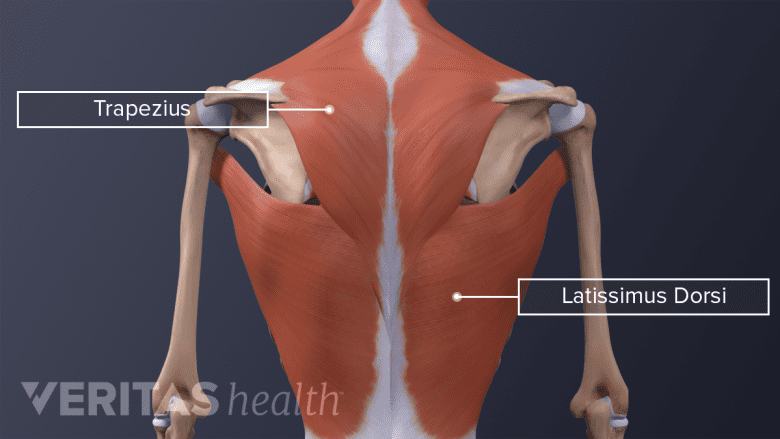
The trapezius and latissimus dorsi span across the neck, shoulder, and back.
Extrinsic muscles originate in the back of the torso and attach to one of the three bones of the shoulder. Extrinsic muscles include:
- The trapezius muscle is a large back muscle with a trapezoid-like shape that covers the shoulder and neck. It connects the vertebral column with the shoulder blade and the collarbone. The trapezius muscle is involved in the internal rotation of the shoulder and the extension of the arm upwards (elevation) and back in a downward direction (depression).17Tiwana MS, Sinkler MA, Bordoni B. Anatomy, Shoulder and Upper Limb, Triceps Muscle. In: StatPearls. StatPearls Publishing; 2023. Accessed July 27, 2023. http://www.ncbi.nlm.nih.gov/books/NBK536996/
- The rhomboid muscles include the rhomboid major and rhomboid minor muscles. These muscles span from the shoulder blade to the vertebral column. The rhomboid muscles contribute to the stability of the shoulder girdle and the movement of the scapula and the upper arm.18Farrell C, Kiel J. Anatomy, Back, Rhomboid Muscles. In: StatPearls. StatPearls Publishing; 2023. Accessed July 27, 2023. http://www.ncbi.nlm.nih.gov/books/NBK534856
- The latissimus dorsi is a broad muscle that occupies the middle and lower back. It is one of the largest muscles in the back and is commonly referred to as “lats.” It originates from the vertebral column, and among its several attachments include the upper arm bone and the shoulder blades. The latissimus dorsi is involved in the upward extension of the arms (elevation) and their sideways movement back toward the midline from a raised position (adduction).19Jeno SH, Varacallo M. Anatomy, Back, Latissimus Dorsi. In: StatPearls. StatPearls Publishing; 2023. Accessed July 27, 2023. http://www.ncbi.nlm.nih.gov/books/NBK448120/
- The levator scapulae muscle arises from the neck vertebrae and attaches to the shoulder blades. It functions in conjunction with the rhomboid and trapezius muscles to elevate the shoulder blades.20Henry JP, Munakomi S. Anatomy, Head and Neck, Levator Scapulae Muscles. In: StatPearls. StatPearls Publishing; 2023. Accessed July 27, 2023. http://www.ncbi.nlm.nih.gov/books/NBK553120
Chest and upper arm muscles
Several muscles in the chest and upper arm also help stabilize and support the shoulder joint.
- The biceps brachii (biceps) are attached to the shoulder blade at two distinct sites. The long head attaches to the glenoid cavity, whereas the short head attaches to the coracoid process. The long head of the biceps is notable for its role in shoulder stability during the elevation of the arms and flexing of the forearms at the elbow.21Tiwana MS, Charlick M, Varacallo M. Anatomy, Shoulder and Upper Limb, Biceps Muscle. In: StatPearls. StatPearls Publishing; 2023. Accessed July 27, 2023. http://www.ncbi.nlm.nih.gov/books/NBK519538/
- The triceps brachii (triceps) has three heads, with one head arising from the shoulder blade and the other two from the upper arm bone. The triceps helps stabilize the upper arm bone during the movement of the upper arms backward from the resting position (extension) and the sideways movement of the arms back towards the midline from a raised position (adduction).17Tiwana MS, Sinkler MA, Bordoni B. Anatomy, Shoulder and Upper Limb, Triceps Muscle. In: StatPearls. StatPearls Publishing; 2023. Accessed July 27, 2023. http://www.ncbi.nlm.nih.gov/books/NBK536996/
- The pectoralis major muscles connect the front walls of the rib cage to the upper arm and the shoulder. The pectoralis major muscle is involved in the sideways movement of the arms from an elevated position back to the midline. In addition, this muscle is also involved in the internal rotation of the shoulder and stabilization of the shoulder blade.22Baig MA, Bordoni B. Anatomy, Shoulder and Upper Limb, Pectoral Muscles. In: StatPearls. StatPearls Publishing; 2023. Accessed July 28, 2023. http://www.ncbi.nlm.nih.gov/books/NBK545241/
- The pectoralis minor is a thin, triangular muscle that lies below the pectoral major. It is involved in the stabilization of the shoulder blades during various movements, and weakness of the pectoralis minor can cause shoulder blade pain.19Jeno SH, Varacallo M. Anatomy, Back, Latissimus Dorsi. In: StatPearls. StatPearls Publishing; 2023. Accessed July 27, 2023. http://www.ncbi.nlm.nih.gov/books/NBK448120/
advertisement
Blood and Nerve Supply of the Shoulder
Blood vessels and nerves travel through the shoulder, providing blood supply and sensation to the shoulder blade.
Blood supply

The brachial artery supplies the shoulder bones and upper arm muscles.
The major artery that supplies blood to the shoulder and the arm region is the subclavian artery.23Miniato MA, Anand P, Varacallo M. Anatomy, Shoulder and Upper Limb, Shoulder. In: StatPearls. StatPearls Publishing; 2023. Accessed July 27, 2023. http://www.ncbi.nlm.nih.gov/books/NBK536933/ The subclavian artery branches near the first rib, with one of the branches entering the shoulder to form the axillary artery. The axillary artery forms several branches, including:
- The subscapular artery
- The superior thoracic artery
- The thoracoacromial artery
- The anterior humeral circumflex artery
- The posterior humeral circumflex artery.
- The brachial artery
The anterior humeral circumflex and posterior humeral circumflex arteries provide blood to the head of the upper arm bone.3Mostafa E, Imonugo O, Varacallo M. Anatomy, Shoulder and Upper Limb, Humerus. In: StatPearls. StatPearls Publishing; 2023. Accessed July 28, 2023. http://www.ncbi.nlm.nih.gov/books/NBK534821/ The brachial artery also supplies blood to the bones in the shoulder and muscles attached to the upper arm bone.
The thyrocervical trunk also arises from the subclavian artery and branches to form the suprascapular artery. The suprascapular artery supplies blood to several muscles, which attach to the shoulder blade and facilitate the movements of the shoulder joints.
Nerves of the shoulder

Nerves transmit signals for arm movement and sensory functions.
Nerves are responsible for providing signals to the brain to move the arms and sense temperature, pain, and touch. Some of the nerves that run through the shoulder are part of and originate in a network called the brachial plexus.3Mostafa E, Imonugo O, Varacallo M. Anatomy, Shoulder and Upper Limb, Humerus. In: StatPearls. StatPearls Publishing; 2023. Accessed July 28, 2023. http://www.ncbi.nlm.nih.gov/books/NBK534821/ The brachial plexus has many nerves that contribute to it, such as:
- The ulnar nerve
- The median nerve
- The radial nerve
- The musculocutaneous nerve
In some people, shoulder pain may be a symptom of a nerve being pinched where it originates - primarily in the cervical spine and the uppermost portion of the thoracic spine. In these cases, the shoulder may feel numb, weak, or have a “pins and needles” feeling.
Nerve blocks—numbing medication that is injected into a group of nerves—may be used to diagnose, prevent, or treat certain types of shoulder pain. In some cases, nerve blocks will be used as a form of regional anesthesia during surgery.
- 1 Chang LR, Anand P, Varacallo M. Anatomy, Shoulder and Upper Limb, Glenohumeral Joint. In: StatPearls. StatPearls Publishing; 2023. Accessed July 27, 2023. http://www.ncbi.nlm.nih.gov/books/NBK537018/
- 2 Wong M, Kiel J. Anatomy, Shoulder and Upper Limb, Acromioclavicular Joint. In: StatPearls. StatPearls Publishing; 2023. Accessed July 28, 2023. http://www.ncbi.nlm.nih.gov/books/NBK499858/
- 3 Mostafa E, Imonugo O, Varacallo M. Anatomy, Shoulder and Upper Limb, Humerus. In: StatPearls. StatPearls Publishing; 2023. Accessed July 28, 2023. http://www.ncbi.nlm.nih.gov/books/NBK534821/
- 4 Faruqi T, Rizvi TJ. Subacromial Bursitis. In: StatPearls [Internet]. Treasure Island (FL): StatPearls Publishing; 2023 Jan. Accessed August 28, 2023. https://www.ncbi.nlm.nih.gov/books/NBK541096/
- 5 Conduah AH, Baker CL, Baker CL. Clinical Management of Scapulothoracic Bursitis and the Snapping Scapula. Sports Health. 2010;2(2):147-155. doi:10.1177/1941738109338359
- 6 Gervasi E, Sebastiani E, Spicuzza A. Multidirectional Shoulder Instability: Arthroscopic Labral Augmentation. Arthrosc Tech. 2017;6(1):e219-e225. doi:10.1016/j.eats.2016.09.025
- 7 Almajed YA, Hall AC, Gillingwater TH, Alashkham A. Anatomical, functional and biomechanical review of the glenoid labrum. J Anat. 2022;240(4):761-771. doi:https://doi.org/10.1111/joa.13582
- 8 Clavert P. Glenoid labrum pathology. Orthop Traumatol Surg Res. 2015;101(1, Supplement):S19-S24. doi:https://doi.org/10.1016/j.otsr.2014.06.028
- 9 Kiel J, Taqi M, Kaiser K. Acromioclavicular Joint Injury. In: StatPearls. StatPearls Publishing; 2023. Accessed July 31, 2023. http://www.ncbi.nlm.nih.gov/books/NBK493188/
- 10 Elzanie A, Varacallo M. Anatomy, Shoulder and Upper Limb, Deltoid Muscle. In: StatPearls. StatPearls Publishing; 2023. Accessed July 27, 2023. http://www.ncbi.nlm.nih.gov/books/NBK537056/
- 11 Syros A, Rizzo MG. Anatomy, Shoulder and Upper Limb, Teres Major Muscle. [Updated 2023 Apr 17]. In: StatPearls [Internet]. Treasure Island (FL): StatPearls Publishing; 2023 Jan-. Available from: https://www.ncbi.nlm.nih.gov/books/NBK580487/
- 12 Varacallo M, El Bitar Y, Mair SD. Rotator Cuff Syndrome. In: StatPearls. StatPearls Publishing; 2023. Accessed July 27, 2023.http://www.ncbi.nlm.nih.gov/books/NBK531506/
- 13 Juneja P, Hubbard JB. Anatomy, Shoulder and Upper Limb, Arm Teres Minor Muscle. In: StatPearls. StatPearls Publishing; 2023. Accessed July 27, 2023. http://www.ncbi.nlm.nih.gov/books/NBK513324/
- 14 Jeno SH, Munjal A, Schindler GS. Anatomy, Shoulder and Upper Limb, Arm Supraspinatus Muscle. In: StatPearls. StatPearls Publishing; 2023. Accessed August 2, 2023. http://www.ncbi.nlm.nih.gov/books/NBK537202/
- 15 Williams JM, Sinkler MA, Obremskey W. Anatomy, Shoulder and Upper Limb, Infraspinatus Muscle. In: StatPearls. StatPearls Publishing; 2023. Accessed July 27, 2023. http://www.ncbi.nlm.nih.gov/books/NBK513255/
- 16 Kellam P, Kahn T, Tashjian RZ. Anatomy of the Subscapularis: A Review. J Shoulder Elb Arthroplasty. 2019;3:2471549219849728. doi:10.1177/2471549219849728
- 17 Tiwana MS, Sinkler MA, Bordoni B. Anatomy, Shoulder and Upper Limb, Triceps Muscle. In: StatPearls. StatPearls Publishing; 2023. Accessed July 27, 2023. http://www.ncbi.nlm.nih.gov/books/NBK536996/
- 18 Farrell C, Kiel J. Anatomy, Back, Rhomboid Muscles. In: StatPearls. StatPearls Publishing; 2023. Accessed July 27, 2023. http://www.ncbi.nlm.nih.gov/books/NBK534856
- 19 Jeno SH, Varacallo M. Anatomy, Back, Latissimus Dorsi. In: StatPearls. StatPearls Publishing; 2023. Accessed July 27, 2023. http://www.ncbi.nlm.nih.gov/books/NBK448120/
- 20 Henry JP, Munakomi S. Anatomy, Head and Neck, Levator Scapulae Muscles. In: StatPearls. StatPearls Publishing; 2023. Accessed July 27, 2023. http://www.ncbi.nlm.nih.gov/books/NBK553120
- 21 Tiwana MS, Charlick M, Varacallo M. Anatomy, Shoulder and Upper Limb, Biceps Muscle. In: StatPearls. StatPearls Publishing; 2023. Accessed July 27, 2023. http://www.ncbi.nlm.nih.gov/books/NBK519538/
- 22 Baig MA, Bordoni B. Anatomy, Shoulder and Upper Limb, Pectoral Muscles. In: StatPearls. StatPearls Publishing; 2023. Accessed July 28, 2023. http://www.ncbi.nlm.nih.gov/books/NBK545241/
- 23 Miniato MA, Anand P, Varacallo M. Anatomy, Shoulder and Upper Limb, Shoulder. In: StatPearls. StatPearls Publishing; 2023. Accessed July 27, 2023. http://www.ncbi.nlm.nih.gov/books/NBK536933/

advertisement